Large submucous uterine fibroid mass
Obiozor AA1, Obiozor CG2
Abstract
Background: Uterine fibroids or leiomyomas, are common benign tumors originating from the smooth muscle cells of the uterus. While typically asymptomatic, large submucous fibroids can cause significant clinical manifestations, including abnormal uterine bleeding, pelvic pain, and reproductive issues. Radiological imaging, such as ultrasound, played a crucial role in the accurate diagnosis and characterization of these fibroids, guiding appropriate clinical management decisions.
Methods: We present a case report of a 28-year-old female with complaints of heavy menstrual bleeding and pelvic discomfort. Clinical evaluation prompted the utilization of radiological imaging techniques to assess the uterine anatomy. Ultrasound was employed to visualize and characterize the large submucous uterine fibroid mass. Imaging findings were correlated with clinical symptoms to formulate an appropriate treatment plan.
Results: ultrasound revealed a large submucous uterine fibroid mass measuring 105.7mm x 75.6mm x 79.6mm in its dimensions. The fibroid was heterogeneous in its echo texture and it is located in the submucosal layer of the uterus. Doppler imaging demonstrated some areas of flow within the fibroid mass. The correlation of imaging findings with clinical symptoms aided in determining the extent of fibroid-related symptoms and guiding further management decisions.
Conclusion: This case report underscores the significance of radiological imaging, particularly ultrasound in diagnosing and characterizing large submucous uterine fibroid masses. Accurate radiological assessment is crucial for determining the appropriate course of clinical management, which may include medical therapy, minimally invasive procedures, or surgical intervention. The collaborative approach between gynecologists and radiologists is pivotal in optimizing patient care for individuals with symptomatic uterine fibroids.
Keywords: Uterine fibroids, radiology, ultrasound, clinical management.
Introduction
Uterine fibroids are benign tumors originating from the smooth muscle cells of the uterine wall, it is a common gynecological problem affecting a significant proportion of women during their reproductive years. Among the diverse presentations of uterine fibroids, submucous fibroids, which develop beneath the inner lining of the uterus, often manifest with distinct clinical symptoms, including abnormal uterine bleeding, abdominal pain and reproductive challenges. This case report deals with a patient harboring a large submucous uterine fibroid mass—and explores the pivotal role of radiological techniques in unraveling its complexities.
The understanding of uterine fibroids has evolved with advancements in diagnostic imaging modalities, like transvaginal ultrasound and magnetic resonance imaging (MRI), computerized axial tomographic scan (CT scan). These tools not only offer precise anatomical localization but also provide detailed insights into the size, composition, and impact on surrounding structures. The aim of the case study is to contribute to the existing body of knowledge surrounding submucous uterine fibroids, shedding light on the challenges posed by large masses and elucidating the diagnostic significance of contemporary radiological approaches.
Given the clinical implications of submucous uterine fibroids, like infertility, abnormal uterine bleeding, abdominal pain, it is essential to arrive at a detailed diagnosis using the right imaging modality. Through this report, we aim to underscore the importance of radiological assessments in facilitating accurate diagnosis and guiding tailored therapeutic interventions. By examining the unique characteristics of a large submucous uterine fibroid mass which was over 10cm in diameter, we also strive to enhance the understanding of clinicians and radiologists alike, fostering improved patient management and outcomes in the realm of women's health.
Case report
This case details the diagnosis of a 28-year-old female that presented with bleeding per vaginum, weakness, and dizziness in a radio-diagnostic center. Clinical examination prompted a comprehensive diagnostic evaluation. Pelvic ultrasound revealed a bulky uterus harboring a large fibroid mass measuring 105.7mm x 75.6mm x 79.6mm and located in the submucous layer. The uterus itself measures 120.7mm x 80.2mm x 87.9mm. There is an anechoic fluid collection within the endometrial cavity. Both adnexae and the abdominal organs were sonographically normal. Emergency myomectomy was done for the patient and patient had a remarkable improvement post-surgery, specimen was sent for histological analysis which came out to be a mass of smooth muscle origin and of benign consistency, diagnosis of uterine fibroid was made which is in consistency with the radiological findings. This case underscores the need for a multidisciplinary approach, involving gynecologists, radiologists, and other specialists. The case highlights the pivotal role of radiological imaging in timely identification, characterization, and management of substantial uterine abnormalities. Such collaborative efforts are essential for optimizing patient care and achieving the best possible outcomes.
Discussion
The presented case report delves into the complexities of diagnosis and management associated with a large submucous uterine fibroid mass, underscoring the crucial role of radiological imaging in guiding patient care. By integrating the ultrasound findings, a comprehensive understanding of the fibroid's size, location, and nature was achieved, influencing treatment decisions and post-treatment strategies. The incidence of submucosal leiomyomas of the uterus is approximately 5-10%.1 Although less common than subserosal and intramural leiomyomas, submucosal myomas are associated with greater morbidity, including dysmenorrhea, menorrhagia, symptomatic anemia requiring blood transfusions, infertility, and an increased risk of early pregnancy complications [1].
The radiological evaluation with ultrasound unveiled specific features of the large submucous uterine fibroid mass. Ultrasound provided initial insights, offering valuable information for treatment planning and ensuring a thorough understanding of the fibroid's characteristics. The comprehensive assessment with ultrasound proved crucial for guiding appropriate interventions.
Large submucous uterine fibroids present diagnostic challenges, demanding a reliance on imaging to complement clinical symptoms. The radiological findings played a pivotal role in establishing an accurate diagnosis, addressing the limitations of symptom-based evaluations. The integration of imaging not only confirmed the presence of the fibroid but also provided crucial details essential for a nuanced understanding of the condition.
The radiological characterization exerted a significant influence on treatment decisions. The size and location of the fibroid played a pivotal role in choosing between conservative management and more invasive interventions, such as myomectomy or hysterectomy. The imaging data played a critical role in tailoring the treatment strategy based on the fibroid's impact on surrounding structures, ensuring a personalized and effective approach to patient care.
Depending on the severity of symptoms, treatment options may be medical or surgical. Medical management includes hormonal therapy such as oral contraceptives, single-agent progesterone suppression, or gonadotrophin-releasing hormone agonists.2 Hormonal treatment is often combined with non-hormonal options that include non-steroidal anti-inflammatory drugs and tranexamic acid.2
This comprehensive approach involves not only addressing the anatomical considerations revealed by radiological imaging but also considering the patient's symptomatology and overall well-being. The integration of medical and surgical interventions, guided by both clinical and radiological assessments, allows for a nuanced and tailored treatment plan that optimizes outcomes while minimizing the impact on the patient's quality of life.
Furthermore, it is crucial to recognize the broader implications of uterine fibroids on women's health. Fibroids can affect fertility,3 influencing reproductive choices and family planning. Additionally, the psychological impact of fibroids on a woman's life should not be overlooked.4 The emotional and mental well-being of the patient is an integral aspect of holistic care, highlighting the importance of a multidisciplinary approach that takes into account both the physical and emotional aspects of managing large submucous uterine fibroid masses.
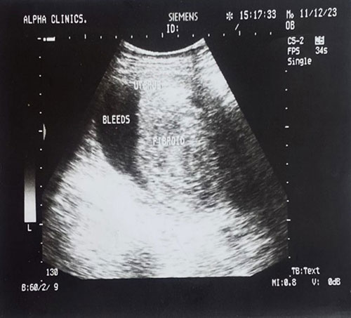
Fig. 1: Ultrasound showing bleeds

Fig. 2: Ultrasound scan of the fibroid
Conclusion
Collaboration between gynecologists, radiologists, and other specialists is essential for a holistic and patient-centered approach in managing large submucous uterine fibroid masses, addressing not only the physical aspects but also the reproductive and psychological dimensions of the patient's well-being. Long-term follow-up remains essential post-treatment, with imaging serving as a key tool in monitoring changes, assessing intervention efficacy, and detecting potential complications or recurrence. Regular follow-up examinations, guided by radiological assessments, are crucial for maintaining patient well-being and addressing emerging issues promptly. The comprehensive insights gained through imaging modalities contribute to informed decision-making, ultimately shaping personalized patient care strategies and ensuring optimal long-term outcomes. In conclusion, this case underscores the integral role of radiological imaging in the diagnosis and management of large submucous uterine fibroid masses, emphasizing its impact on treatment decisions and the overall trajectory of patient care.
References
- Liang B, Xie YG, Xu XP, Hu CH. Oncol Lett. Diagnosis and treatment of submucous myoma of the uterus with interventional ultrasound. 2018;15:6189–6194.
- Sohn GS, Cho S, Kim YM, Cho CH, Kim MR, Lee SR. Current medical treatment of uterine fibroids. Obstet Gynecol Sci. 2018;61:192–201
- Whynott RM, Vaught KCC, Segars JH. The effect of uterine fibroids on infertility: a systematic review. Semin Reprod Med. 2017;35(6):523–32.
- Zimmermann A. Prevalence, symptoms and management of uterine fibroids: an international internet-based survey of 21,746 women. BMC Women Health. 2012;12:6.