Effect of periodic exposure to formaldehyde in the anatomy laboratory on some haematological indices in male Wistar rats
Ebojele FO, Iyawe VI
Abstract
Background: The effect of periodic exposure to formaldehyde on some haematological indices in male Wistar rat was studied.
Materials and Methods: The Wistar rats were divided into three groups A,B and C with 5 animals in each group. Group A served as control with nil exposure while groups B and C were the test groups with 5 months exposure on non-dissection days and dissection days respectively. Formaldehyde air level was measured both at experimental and control sites. Some haematological parameters measured include Red blood cell count, Haemoglobin concentration, White blood cell count: total and differential, and platelet count. Statistical analysis was done using Graph Pad Prism version 5.0. Results were presented as Mean ± SEM. Analysis of Variance was used to compare the means of test and control values while post hoc test was done using Student Newman Keul’s test and a P-value of less than 0.05 was considered as statistically significant.
Results: Results revealed significant increase in formaldehyde air level in the dissection hall. There was significant increase in total white blood cell count when compared with control while red blood cell count, haemoglobin concentration and platelet count were not significant.
Conclusion: It was therefore concluded that periodic exposure to formaldehyde in the Anatomy laboratory may have no effect on most haematological indices in Wistar rats although the white blood cells may be affected.
Keywords: periodic, formaldehyde, haematological, Wistar rats.
Introduction
Formaldehyde which is a known chemical fixative used for preserving dead bodies (cadavers) has been reported to produce some undesirable effects especially in the respiratory system in humans.1,2 Available studies on acute occupational exposure in humans revealed clinical symptoms like skin irritation, eye soreness, nose irritation, throat irritation and rhinorrhea as well as reduction in pulmonary function parameters like Forced Vital Capacity (FVC), Forced Expiratory Volume in one second (FEV1), Peak Expiratory Flow Rate (PEFR) and FEV1/FVC ratio.3,4,5,6 Chronic exposure studies done in some medical schools in India reported decrease in pulmonary function following exposure to formaldehyde.7,8,9 Some animal studies have reported oxidative stress in the liver and evidence of lipid peroxidation for exposures as high as 8 part per million.10,11 Some researchers have tried to look at possible effects of formaldehyde on the central nervous system.12,13,14 as well as the reproductive system.15,16 Atmospheric levels of formaldehyde has been reported to be higher in the Gross Anatomy laboratory when compared to other laboratories in a tertiary institution where medical students are trained.17 Medical Students of a tertiary institution where this present study was carried out are usually exposed to the Anatomy laboratory where they carry out dissection for eight hours every week, that is, twice a week for a duration of four hours each. Will this periodic weekly exposure to the Anatomy laboratory have any effect on the haematological indices of the Medical Students? This periodic exposure to the Anatomy laboratory was mimicked in Wistar rats and they were exposed for five months to see if there will be any adverse effect of formaldehyde on some haematological indices and this formed the basis of the present study.
Materials and methods
Experimental animals
Fifteen male Wistar rats of comparable age weighing between 180-220g were procured from the animal house of the Department of Anatomy, University of Benin. The animals were kept in plastic cages with wire mesh floor and allowed to acclimatize for a period of two weeks on normal feeds and water before the commencement of the experiments. Animal management and experimental protocols were carried out in accordance with the recommendations of the 1996 Guide for the Care and Use of Laboratory Animals.18
Animal grouping
The rats were divided into three groups (A, B and C) with five animals in each group. Group A served as the control with nil exposure while groups B and C served as the test group. Group B animals were exposed to formaldehyde in the dissection room on non-dissection days for eight hours per week for a duration of five months while Group C animals were exposed to formaldehyde in the dissection room on dissection days for eight hours per week which was also equivalent to the period medical students spend in the anatomy laboratory during dissection and this was also carried out for a duration of five months.
Measurements of formaldehyde air level
Formaldehyde air level was measured using Formaldehyde Gas Meter (EXTECH Fm200). The meter is automated, calibrated and has an external probe that detects the atmospheric levels of formaldehyde. Five measurements were taken on five different occasions at the control site and in the dissection hall on dissection days and non-dissection days and the average was calculated and taken as the air exposure level. Within the dissection hall, the measurements were taken around the dissection table to get an idea of the personal exposure, and three meters away from the dissection table to get an idea of the area exposure. Measurements were also taken at the different corners of the laboratory. The formaldehyde air levels were measured in part per million (ppm). The meter also gave measurement of the room temperature and the relative humidity.
Sample collection and analysis
Blood samples from the animals were collected through cardiac puncture as described by D’Armour et al.19 The blood samples were transferred into sodium EDTA and sodium citrate containers for haematological analysis. The blood samples collected into the sodium EDTA container was for determination of red cell count (RBC), haemoglobin concentration, total white cell count and its differentials using automated haematology system (Diatron® Abacus junior haematology analyzer), while samples collected into sodium citrate container was for platelet determination.
Statistical analysis
Statistical analysis was done using Graph pad prism version 5.0. Results was presented as Mean ± SEM. Analysis of Variance was used to compare the means of test and control values while post hoc test was done using Student Newman Keul’s test and a p-value of less than 0.05 was considered as statistically significant.
Results
Table 1: Formaldehyde air level, room temperature and relative humidity of control site and
dissection room
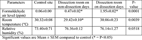
Table 2: Some haematological indices of male Wistar Rats following periodic exposure to
formaldehyde in the Anatomy laboratory
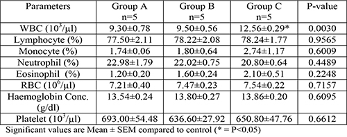
Formaldehyde air level of control site and dissection room is shown in Table 1. There was a significant increase (p<0.05) in formaldehyde air level in the dissection room both on the dissection days and non-dissection days. Total white blood cell count was significantly increased in group C (Table 2) when compared with the control, while Red blood cell count, Haemoglobin concentration and Platelet count showed no significant difference.
Discussion
The atmospheric level of formaldehyde in the Anatomy laboratory measured in this present study was 1.95 part per million on dissection days and 0.47 part per million on non-dissection days (Table 1). This is a reflection of the amount of formaldehyde that the medical students are exposed to each week when they come into the Anatomy laboratory to carry out dissection. When Wistar rats were exposed to this same amount of formaldehyde that the medical students are exposed to we saw a significant increase in Total white blood cell count among the animals exposed on dissection days. Here formaldehyde appears to be having a proliferative effect on the white blood cells resulting in leucocytosis which may be an attempt to counter the effect of formaldehyde. However, Red blood cell count, Haemoglobin concentration, and platelet count were not affected at this exposure level. This therefore suggests that periodic exposure to formaldehyde in the Anatomy laboratory may not have adverse effect on some haematological indices in Wistar rats. In some studies carried out in rats formaldehyde was reported to produce oxidative stress in the liver.10,11 The authors also reported evidence of lipid peroxidation among rats exposed to as high as 8 part per million of formaldehyde. In this present study, air exposure level to formaldehyde for group B rats exposed on non-dissection days was 0.47ppm while that of group C rats exposed on dissection days was 1.95ppm. The exposure level to formaldehyde in present study is not up to the level that produced oxidative stress and lipid peroxidation in the study on rats earlier cited. Recall that this study carried out on wistar rats actually mimicked medical students’ attendance at the Anatomy laboratory for the purpose of dissection during the course of their training. Since most haematological indices that were measured in Wistar rats were not affected, is it possible that haematological indices may not also be affected among medical students who are exposed to the same amount of formaldehyde in the Anatomy laboratory? Further studies among medical students with regard to haematological indices following exposure to formaldehyde is required in order to substantiate these observations in Wistar rats.
Conclusion
Periodic exposure to formaldehyde in the Anatomy laboratory may not have any adverse effect on some haemotological indices in Wistar rats.
References
- Farooqui M.Y.H. Formaldehyde. J Appl Toxicol 1983; 3: 264-265.
- Mathur N, Rastogi S.K. Respiratory effects due to occupational exposure to formaldehyde; systematic review with meta analysis. Indian J Occup and Environ Med 2007; 11: 26-31.
- Khamgaonkar M.B, Fulare M.B. Pulmonary effects of formaldehyde exposure - an environmental epidemiological study. Indian J Chest Dis Allied Sci 1991; 33: 9-13.
- Kim H, Kim Y, Cho S. Formaldehyde exposure levels and serum antibodies to formaldehyde-human serum albumin of Korean medical students. Arch Environ Health 1999; 54(2): 115-18.
- Pourmahabadian M, Azam K, Ghasemkhani M. Pulmonary function study between formaldehyde exposed and non-exposed staffs at some of the Tehran educational hospitals. J Med Sci 2006; 6(4): 621-25.
- Wei C.N, Harada K, Ohmori S. Subjective symptoms of medical students exposed to formaldehyde during a gross anatomy dissection course. Int J Immunopathol Pharmacol 2007; 20(2): 23-25.
- Patil P, Hulke S.M, Thakare A. Effect of formalin on pulmonary function: a nine months longitudinal study. Research J of Pharmaceutical Biol and Chem Sci 2012; 3(1): 211-216.
- Neginhal R, Herur A, Chinagudi S, Rairam G.B, Kolagi S, Ambi U. Cardiorespiratory effects of acute exposure to formaldehyde in gross anatomy laboratory in medical students- A comparative study. Medica Innovatica. 2013; 2(1): 32-35.
- Shrivastava A, Saxena Y. Effect of formalin vapour on pulmonary function of medical students in anatomy dissection hall over a period of one year. Indian J Physiol Pharmacol 2013; 57(3): 255-60.
- Petushok N. Activity of glutathione-related enzymes in rat tissues after formaldehyde exposure. Curr Top Biophys 2000; 24(2): 167-169.
- Sogut S, Songur A, Ozen O. Does the subacute (4-week) exposure to formaldehyde inhalation lead to oxidant/antioxidant imbalance in rat liver? Eur J Gen Med 2004; 1(3): 26-32.
- Usanmaz S, Akarsu E, Vural N. Neurotoxic effects of acute and subacute formaldehyde exposure in mice. Environ Toxicol Pharmacol 2002; 11(2): 93-100.
- Aslan H, Songur A, Tunc T. Effects of formaldehyde exposure on granule cell number and volume of dentate gyrus: A histopathological and sterological study. Brain Res 2006; 1122(1): 191-200.
- Sarsilmaz M, Kaplan S, Songu A. Effects of postnatal formaldehyde exposure on pyramidal cell number, volume of cell layer in hippocampus and hemisphere in the rat: A stereological study. Brain Res 2007; 1145: 157-167.
- Ozen O.A, Akpolat N, Songur A. Effect of formaldehyde inhalation on Hsp70 in seminiferous tubules of rat testes: An immunohistochemical study. Toxicol Ind Health 2005; 21(10): 249-254.
- Zhou D, Qiu S, Zhang J. The protective effect of vitamin E against oxidative damage caused by formaldehyde in the testes of adult rats. Asian J Androl 2006; 8(5): 584-585.
- Ebojele F.O, Iyawe V.I. Laboratory atmospheric levels of formaldehyde in selected laboratories used by medical students in a tertiary institution in Edo State, Nigeria. J Appl Sci Environ Manage 2021; 25(10): 1747-1750.
- Clark J.D, Gebhart G.F, Gonder J.C, Keeling M.E, Kohn D.F. The 1996 Guide for the Care and Use of Laboratory Animals. ILAR Journal 1997; 38(1): 41-48.
- D’Armour F.E, Blood F.R, Belden D.A. The manual for laboratory work in mammalian physiology. 3rd Edition. University of Chicago Press, Illinois Chicago 1965; Pp4-6.