Prepatellar tumoral calcinosis mimicking soft tissue sarcoma: A case report and review of literature
Dim EM1, Ubaha A2, Usendiah IB2, Dim UME3, Oforjigha-Dim CW4, Essien U2
Abstract
Tumoral calcinosis is a rare, painless tumour-like mass often seen as soft tissue calcifications around joints in adolescents and young adults. The occurrence of this rare entity in the middle aged or elderly population is also rare. The pathology may mimick sarcomatous lesions or granulomatous diseases. A high index of suspicion, imaging and laboratory studies, including histopathological examinations, are needed to make a diagnosis. A 59-year old Nigerian woman that complained of a progressive and painless anterior knee mass, which turned out to be a tumoral calcinosis, is presented.
Introduction
Tumoral calcinosis (TC) is a rare lesion characterized by deposition of calcium salts in the soft tissue around the joints, with resultant local symptoms and limitation of movements. It was first described by Duret and the term tumoral calcinosis was coined by Inclan.2,3 It is also known as Teutsecherlaender disease (following studies carried out by Teutsecherlaender between 1930 and 1950), calcifying bursitis, and lipocalcino granulomatosis.4 Swellings are usually found in large joints of the elbow, hip, and shoulder though occasional cases of small joints have been reported.5,6,7 It is seen mostly in adolescents and young adults, although some variants affecting children have been documented.5,8 It is classified by Smack into a primary type, which may arise de novo, or a secondary type. Familial variants have been reported that arise due to the mutation of genes like GaINAc-transferase3 (GALNT3) and fibroblastic growth factor 23 (FGF23).
Patients’ evaluation is clinical, supported with imaging studies and laboratory investigations, including histopathological studies. Surgical excision is the treatment of choice. The aim of this work is to report a case of calcified prepatellar bursitis, mimicking a soft tissue sarcoma, in a 59-year old Nigerian woman, who complained of a progressive, painless, anterior knee swelling of over 12 years prior to presentation.
Case Report
A 59-year old Nigerian woman, who complained of an anterior right knee swelling of 12 years duration, was referred to the orthopaedic clinic with an initial suspicion of a malignant disease involving the knee. The swelling was painless, progressively increased in size, itchy on the overlying skin surface, and occasional discharge of pus from two foci on the swelling. There was a prior history of minor domestic trauma to the knee, just before the onset of swelling, when she hit the knee against an object. There was an associated difficulty with kneeling on the right knee. There was no history of fever or weight loss. The past medical history was not remarkable.
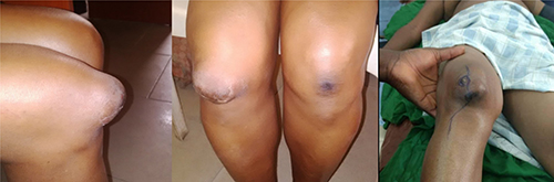
Figure 1: Different views of the anterior knee swelling
Physical examination showed a 6cm x 6cm anterior knee mass, located between the lower pole of the patella and tibia tubercle. It was hard in consistency, non-tender, and without differential warmth. It was attached to the overlying skin and free from the lower pole of the patella. There was an area of cellulitis, scarring and hyperpigmentation, with serosanguinous discharge from two tiny foci. The range of motion of the knee was normal. There was no regional lymphadenopathy (figure 1).
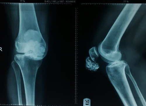
Figure 2: Plain radiographs of the right knee, showing calcific densities in the soft tissue close to the lower pole of the patella.
A plain radiograph of the right knee (anteroposterior and lateral views) showed a mottled hyperdense lesion in the soft tissue plain of the anterior knee joint below the lower pole of the patella (figure 2). The renal function tests, serum calcium and phosphate levels were normal. The serum uric acid level was elevated. The complete blood count showed slight elevation the lymphocytes and the erythrocyte sedimentation rate, probably as a result of a chronic inflammatory process. Fine Needle Aspiration Cytology (FNAC) showed inflammatory cells, comprising mainly polymorphs and lymphocytes.
Figure 3: Intraoperative photograph and the excised tissue
The patient was treated with allopurinol for the background hyperuricaemia, and excision biopsy of the knee mass was done following basic oncology rules (Figure 3), and specimen subjected to pathological examination. The macroscopic findings showed the dimensions of the mass as 5.0cm x 4.0cm x 1.0cm and the cut surface was nodular and tan-yellow in appearance. Microscopy showed nodules of amorphous calcified material separated by bands of dense fibrous tissue with foci of chronic inflammatory cells. The features were consistent with tumoral calcinosis. The patient did well postoperatively, attended a few follow up clinic sessions before she was lost to follow-up after 3 months of out-patient visits.
Discussion and literature review
Tumoral calcinosis (TC) is a rare clinical and histopathological entity resembling neoplasm, and characterized by deposition of calcium phosphate and hydroxyapatite (Ca10(PO4)6(OH)2) salts in different peri-articular soft tissue regions.1 In the index patient, the referral team suspected soft tissue sarcoma as the initial working diagnosis, and patient was referred for a basic musculoskeletal oncology assessment and care. TC is seen in young adults and adolescents as a painless, firm, tumour-like mass located around the joint, especially large joints, which may lead to limitation of joint function.5 Although common in this age group, it is important to note that it can occur in any age like the index patient, who was 59 years. There has been documentation of occurrence in the elderly.9
The Joints commonly affected include the elbow, hip, shoulder, ankle, and wrist.5 Also, there are reported cases occurring in the spine, temporomandibular joint, metacarpal, metatarsal, and popliteal space.5,7 However, the knee was the site of the mass in the index patient. This shows that it can occur in periarticular tissues around the joint as well.
Tumoral calcinosis was first coined by Inclan et al., in 1943.3 He described large juxta-articular lobular calcific masses without a visceral lesion.3 These calcifications were seen in patients with normal serum calcium and phosphorus levels with characteristic pathological features of multiple cysts filled with calcified deposits lined by histiocytes, giant cells, and xanthematous histiocytes.3 Smack et al., in 1996 classified TC into primary and secondary types.10 The primary type is subdivided into primary normo-phosphataemic (sporadic) and primary hyper-phosphatemic (familial) tumoral calcinosis. The hallmark of primary normo-phosphatemic tumoral calcinosis is normal blood calcium and phosphate. This description fits the index case. It is the most common type, occurring before the second decade of life (but the index case occurred at the end of the 5th decade), and is seen in tropical and subtropical regions.10 There is a defect in the sterile α motif domain-containing protein (SAMD9). There are hypotheses that SAMD9 may be a downstream target of tumour necrosis factor -α signalling and, therefore, mediates pro-apoptotic signals activating counter-regulatory anti-inflammatory activities. SMAD9 is also involved in dystrophic calcinosis, a common form of extraosseous calcification. In the primary hyperphosphatemic type there is normal blood calcium with high phosphate levels.12 It occurs in the first and second decades of life with predominance in the black population.12 The primary hyperphosphataemic type is due to decreased urinary phosphate excretion which is a result of recessive mutation in GALNT3, an enzyme that initiates O-linked glycosylation and the α klotho gene. The mutation also leads to the accumulation of FGF23 which is a phosphaturic hormone. This results in uncontrolled production of 1, 25 dihydroxyvitamin D leading to increased reabsorption of phosphate from the kidney and intestine.13 The classifications, although widely propagated in literature, is controversial because histology is similar in the three groups.14
Aetiopathogenesis described in literature involves a common pathway that results in the formation of the characteristic lesion. This occurs because of minimal repetitive trauma or chronic pressure on the tissue which results in haemorrhage in periarticular tissues initiating a foamy histiocytic response.14 We believe that this explanation applied in the aetiopathogenesis of the index case. The calcified debris fills the loculi leading to bone formation with the arrest of bursae formation (neo bursae).14 Decrease in collagenolytic activity results in fibrosis surrounding the tumoral calcinosis lesion. The secondary type is often associated with chronic renal failure and hyperparathyroidism.12,13 The index patient was likely the sporadic type because the calcium, phosphate, and renal parameters were within normal range. The increased swelling over the years may be related to repeated minimal trauma following kneeling down to pray as this was her custom.
Laboratory investigations to aid diagnosis of TC may include serum calcium, phosphate, alkaline phosphate, vitamin D, parathyroid hormone, erythrocyte sedimentation rate, rheumatoid factor, and anti-cyclic citrullinated peptide.13,14 Some of these tests were conducted on the index case.
Plain radiographs are often diagnostic. It shows multiple areas of well-circumscribed nodular masses with fibrous septate which gives a “cobblestone” or “chicken-wire” appearance.15 Computed tomography scan shows “sedimentation sign” due to mineral portion pooling dependently causing a fluid calcium level. Magnetic resonance imaging T1 weighted is homogenous low signal intensity, while T2 shows mixed signal intensities or appears homogenous with high signal intensity.15
Symptomatic treatment is done to correct any imbalance of calcium or phosphate. Medical treatment which involves the use of steroids, and radiation therapy have been discussed by authors, but the results are poor. Needle aspiration has been documented to help relieve symptoms. However, surgical excision of the mass with phosphate deprivation in the hyperphosphatemic type is done for long-term control. Complete surgical excision of mass prevents recurrence. Regression of the mass occurs but it is a rare event.8,15
Conclusion
Tumoral calcinosis should be considered a differential diagnosis of soft tissue calcification. It is a rare pathology; therefore, a high index of suspicion and a multidisciplinary approach is required to make a diagnosis. The patient must be properly evaluated clinically and should undergo various diagnostic and imaging modalities. Complete surgical removal to prevent recurrence is the best approach. Long-term follow-up is important because of its high rate of recurrence.
References
- Lai L, Hsiao M, Wu C, Ozcakar L. Big gain, no pain: Tumoral Calcinosis. The American Journal of Medicine. 2018;1(131):45-7.
- Duret MH. Tumours multiples et singulieres des bourses sereuses (endotheliomes, peutetre d’origine parasitaire) Bull Mem Soc Anat Paris. 1899;74:725-33
- Inclan A, Leon P, Camejo MP. Tumoral calcinosis. Journal of American Medical Association 1943;121(7):490-95.
- Barriere H, Welin J, Lenne Y, Visset J, Viger P. [Teutschlaender lipo-calcino-granulomatosis or tumoral calcinosis of Inclan (author’s transl)] Ann Dermatol Venerol. 1977;104(2):136-40.
- Ibrahim F, Mahmod S. Review of tumoral calcinosis: A rare clinic-pathological entity. World Journal of Clinical Cases. 2014;2(9): 409-14.
- Durant DM, Riley LH, Burger PC, Mccarthy EF. Tumoral calcinosis of the spine: a study of 21 cases. Spine (Philia Pa 1976). 2001;26: 1673-679.
- Hammert WC, Lindsay LR. Tumoral calcinosis-or is it? A case report and review. Hand (New York). 2009;2013:119-22.
- Niali DM, Fogarty EE, Dowling FE, Moore DP. Spontaneous regression of tumoral calcinosis in an infant; a case report. Journal of Paediatric Surgery 1998;33:1429-431.
- Croock AD, Silver RM. Tumoral calcinosis presenting as adhesive capsulitis: case report and literature review. Journal of American College of Rheumatology. 1987;30(4)455-58.
- Smack D, Norton SA, Fitzpatrick JE. Proposal for a pathogenesis-based classification of tumoral calcinosis. Int J Dermatol. 1996;35(4):265-71.
- Hershkovitz D, Gross Y, Nahum S, Yehezkel S, Sang O, Uitto J, Sprecher E. Functional characterization of SAMOD9, a protein deficient in normophosphatemic familial tumoral calcinosis. J Invest Dermatol. 2011;131(3):662-9. doi 10.1038/jid.2010.387.
- Cheftez I, Ben AD, Browning S, et al. Normophosphatemic familial tumoral calcinosis is caused by deleterious mutations in SAMD9, encoding a TNF-alpha responsive protein. J Invest Dermatol. 2008;2013:1423-
- Farrow EG, Imel EA, White KE. Miscellaneous non-inflammatory musculoskeletal conditions. Hyperphosphataemic familial tumoral calcinosis (FGF23, GALNT3 and αklotho). Best Pract Res Clin Rheumatol 2011;2013:735-47
- Slavin RE, Wen J, Barmada A. Tumoral calcinosis—a pathogenetic overview: a histological and ultrastructural study with a report of two new cases, one in infancy. Int J Surg Pathol. 2012;20(5):462-73. doi: 10.1177/1066896912444925.
- Martinez S, Vogler III JB, Harrelson JM, Lyles KW. Imaging of tumoral calcinosis: new observations. Radiology 1990;174:215.