Sonographic Measurement of the Liver in the healthy adult Nigerian Population
Obiozor AA
Abstract
Background: This study is on sonographic measurement of the liver in the healthy adult Nigerian population. The objective of the study is to determine normal lengths of the liver and to correlate the measurements with the sex, age, height, weight and body mass index, in the adult Nigerian population.
Materials and Methods: A sample size of five hundred (500) was used for the study and the individuals were not less than twenty (20) years as at the time of data collection. A questionnaire was used for inclusion and exclusion criteria. Consent was obtained from the individuals before the study and strict confidentiality was maintained. Descriptive and inferential statistics were deployed to realize the objectives.
Results: The mean length (L1) of the liver was 117.2±12.5mm for males and 119.2±13.3mm for females with P values of 0.087 which is not significant. More so, the mean length (L2) of the liver was 133.1±10.1mm for males and 134.5±10.8 for females with P values of 0.133 which is not significant. There is positive correlation of the liver lengths (L1)with age P values 0.000, height P values 0.006 and width with P values 0.019.
Conclusion: Liver size is affected by many variables such as age and weight in both genders. Liver length decreased with age, and size of the liver in males was greater than in the females. Also, there is a significant positive correlation with liver dimensions and the variables studied.
Keywords: Liver, Sonography, nomogram, measurement
Introduction
The liver weighs approximately 1500g and is located in the right upper quadrant of the abdomen; it is also the largest abdominal organ in the human body.1 It performs various functions such as metabolism of carbohydrates, proteins and lipid. It produces bile that helps in the emulsification of fats, and also helps in detoxification of drugs and in the storage of vitamins.2
Brief Anatomy of the liver
The liver is the largest organ in the body.1-4 It is essential for life, performing several complex functions in the human body.2,5,6 It is situated at the right upper quadrant of the abdomen underneath the lower ribs. It extends approximately from the 6th rib down the right costal margin. This makes it difficult to palpate the whole organ in clinical examination.3 Clinically, the liver size is measured by palpation and percussion of the right hypochondrium. Measurements are made in centimeters below the costal margin.3,5
A normal sized liver may be felt below the right costal margin, this does not mean that it is enlarged.5 Therefore this method is very subjective in the determination of liver size.7,8 A more accurate, reliable and reproducible method of determining the liver size is by imaging.
Techniques such as ultrasonography, magnetic resonance imaging, computerized tomography and plain film radiography have been used.7,8 However ultrasound is chosen because it is non ionizing, non-invasive, cheap, reliable and reproducible.3 Ultrasonography is the use of high frequency sound waves of about 2-15MHz to visualize both deep and superficial organs of the body. It involves the transmission of high frequency sound and relies on the reception of the reflected sound waves (echoes) via a transducer. Transmitted sounds are displayed as real time images in the B- mode. Ultrasound is the imaging modality of choice in investigating the liver and the biliary system because apart from being non-ionizing, it can differentiate between solid and cystic lesion of the liver.3
Various dimensions of the liver viz the cranio-caudal length, antero-posterior, and transverse diameters of the liver can be measured using ultrasound.9 These measurements could be used to calculate the volume of the liver.9
Ultrasonography also has the advantage of evaluating other abdominal organs at the same time. When scanning, the liver, the biliary system can also be assessed. The echo-texture during the examination of the liver is compared, with that of the kidneys, pancreas, and spleen. Anthropometrically the liver size is said to be influenced by height, weight, and body mass index.9
Alcohol consumption, pathological conditions like cirrhosis (of any cause), tumours (e.g. hepatoma, lymphoma, metastasis); malaria, miliary tuberculosis, amyloidosis, syphilis, storage disorders, congestive cardiac failure and others may also cause an enlarged liver.4
A diseased liver may be enlarged or shrunken depending on the nature and chronicity of the disease.3 An enlarged liver usually does so by extending downwards (caudally). Hence, measuring the cranio caudal length (span) of the liver at a reference point (e.g. mid-clavicular line) will aid in detecting hepatomegaly.9 Since a diseased liver could be enlarged or shrunken, it is therefore important that a standard range of values for normal liver size is established in our own environment, hence the purpose of this study.
Methodology
This study is on sonographic measurement of the liver in the healthy adult Nigerian population in Enugu State, subjects were recruited amongst undergraduates in higher institutions in Enugu that came for their routine medical examination in UNTH Enugu. Questionnaire was used for inclusion and exclusion criteria. The objective of the study is to determine the nomogram for the liver and to correlate the measurements with the sex, age, height, weight and body mass index, in the adult Nigerian population.
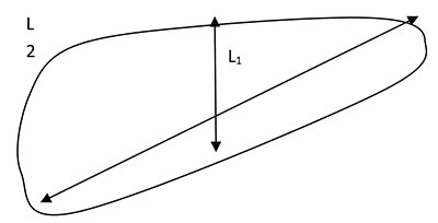
Fig. 1: Sketch of the landmark for sonographic measurement of the liver
A sample size of five hundred (500) was used for the study and the individuals were not less than twenty (20) years as at the time of data collection. A questionnaire was used for inclusion and exclusion criteria. The inclusion criteria were, subjects should not be less than 20yrs of age, no history of hepatitis or any debilitating illness as at the time of study, while exclusion criteria were diabetes, hypertension, haemoglobinopathies, malignancies, etc. Consent was obtained from the individuals. Hands were placed under the head to give free access to the right hypochondrial region. Measurements were taken from the upper margin of the liver along the mid clavicular line to the lower margin of the liver (L1) and diagonally from the left upper most part of the liver margin to the right inferior margin of the liver (L2)
The sample size was estimated using the following formula according to Araoye10
N = Z2S2 / d2
N = Sample size, Z = the standard normal deviate usually set at 1.96, which corresponds to the 95 percent confidence interval, S = Best estimate of organ dimension variance from literature 0.56, d = degree of accuracy desired usually set at 0.05
Thus N = (1.96)2 x (0.56)2 / 0.052
= 3.8416 x 0.3136 / 0.0025 = 481
The minimum sample is 481 subjects however allowing for 5% attrition and non-compliance rate;11 the minimum sample size becomes 506 subjects.
The data was analyzed using SPSS statistical package version 21.0. The mean, median, variance, standard deviation of the liver sizes were evaluated and correlated with the age, sex, weight, height and body mass index of the subjects.
Approval for the study was sought and obtained from the Research and Ethics Committee of University of Nigeria Teaching Hospital.
Ultrasonography was chosen because it is devoid of radiation, non-invasive, real-time and gives accurate measurements.
Results
Table 1: Bio-data of study participants
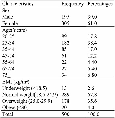
Table 2: Liver dimension according to sex
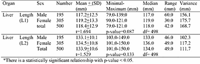
Table 3: Liver mid-clavicular length (L1) by age and sex
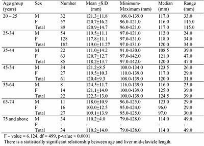
Table 4: Longest (inferior-superior) diagonal length of the liver (L2) by age and sex
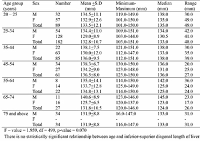
Table 5: Liver dimension by body mass index
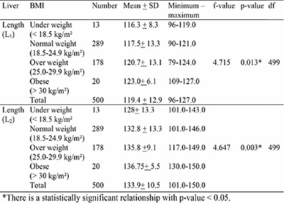
Table 6: Correlation analysis of liver dimension with age, BMI, heigth and weight of subjects.
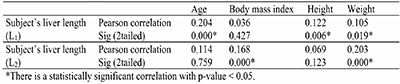
The mean length (L1) of the liver was 117.2±12.5mm for males and 119.2±13.3mm for females with p-values of 0.087 which is not significant. More so, the mean length (L2) of the liver was 133.1±10.1mm for males and 134.5±10.8 for females with p-values of 0.133 which is not significant. There is positive correlation of the liver length (L1) with age p-values 0.000, height p-values 0.006 and width with p-values 0.019. There is positive correlation of the liver length (L2) in the sex but the body mass index p-values 0.000, width with p-values 0.000 but not with age p-values 0.759 and height p-values 0.123.
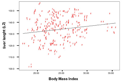
Figure 2: Scatter diagram of liver length (L2) by body mass index
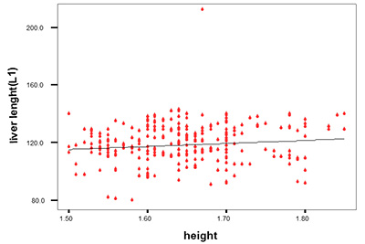
Figure 3: Scatter diagram of liver length (L2) by body mass index
Discussion
The total number of subjects studied was 500; Males were 195 representing 39% of the population, while 305 females representing 61% of the population were studied.
This study shows that the mean (cranio-caudal) mid-clavicular length of the liver (L1) was 117.2±4.5mm and 119.2±13.3mm for males and females respectively, while the range was 79.0-139.0mm for males and 90.0-121.0mm for females. the mean longest (inferior-superior) diagonal length of the liver (L2) was 133.1±10.1mm and 134.5±10.8mm for males and females respectively with a range of 103.0-149.0mm for males and 101.0-150.0mm for females. These measurements are in agreement with the defined limits of the normal liver which was 150mm by Obajimi et al12 in their abdominal ultrasonography in HIV/AIDS patients in the south-western Nigeria. There is a significant correlation of the liver length (L1) with age p values 0.000, height p-values 0.006 and weight p-values 0.019, but not with body mass index. As these parameters increases, the liver length also increases. It is also noted that at age 75 years and above, the liver length decreased with age. It has also shown that the size of the liver in males was greater than in the females. The range of the liver length (L1) from the study was 79.0-139.0mm and 90.0-121.0mm for males and females respectively, while the range of L2 was 103.0-149.0mm and 101.0-150.0mm for males and females respectively. The study also suggests that measurement of the liver length greater than 150 is abnormal.
Conclusion
Liver size is affected by many variables such as age and weight in both genders. Liver length decreased with age, and size of the liver in males was greater than in the females. Also, there is a significant positive correlation with liver dimensions and the variables studied. This study provides a reference value to evaluate liver sizes. Liver dimensions were significantly higher in males compared to females.
References
- Hacer Y, Mechmet S, Erol H. Sonograhpic Measurement of the Liver, Spleen and Kidney in Healthy Term Newborns. America Journal of Radiology 2003; 180: 35 – 40.
- Barrett, K. E. Ganong’s review of medical physiology (24 th ed.). New york: McGraw-Hill Medical. 2012
- Singh, T. Clinical and Sonographic Estimation of Liver Span in Normal Healthy Adults. International Journal of Medical Research & Health Science, 2017. 6(1),
94-97. - Childs, J. T. Methods of Determining the Size of the Adult Liver Using 2D Ultrasound. Journal of Diagnostic Medical Sonography, 2014. 30(6), 296-306.
- Michael Swash ed. Hutchison’s clinical method 21st edition. Edinburgh. WB Saunders Harcourt Publishers 2002: 141 – 162.
- Marc Ghany, Jay H. Hoofnagle In Eugene Braunwald (ed). Liver and biliary disease. Harrison’s principles of internal medicine 15th edition. New York. The McGraw – Hill Company Inc. 2001: 17017 – 1714.
- Zoli M, Magalotti D, Grimaldi M, Gueli G, Marchesini G. Pisi E. Physical examination of the liver: Is it still worth it? Am J Gastroenterol 1995; 90(9): 1428 – 32.
- Joshi R. Singh A, Jajoo N, Pai M, Kalantri SP. Accuracy and reliability of palpation and percussion for detecting hepatomegaly: a rural hospital-based study. Indian J Gastroenterol 2004, 23(5): 163 – 4.
- Kawamura DM, Carr – Hoefer C In. Kawamura DM(ed). Diagnostic Medical Sonography A guide to clinical practice. Abdomen and Superficial structures. 2nd edition Lippincott Raven 1997; 263 – 288.
- Araoye M. O. (2004) Sample size determination. In Research Methodology with Statistics for health and social science. Araoye M.O. (ed). Nathadex Publishers Ilorin. 2004; P.115 – 129.
- Afolabi B. A companion of medical statistics, Ibadan; Ibipress & Publishing CO. 2006;161
- Obajimi M, Atalabi O. Abdominal ultrasonography in HIV/AIDS patients in the south-western Nigeria, BMC Medical Imaging.2008; 8:5.