Computed tomographic evaluation of head injury patients in Umuahia, Abia State Nigeria
Obiozor AA, Imo A, Aka CK
Abstract
Background: Trauma to the scalp, skull and brain is defined as head injury. Head injury is a silent killer, especially in patients that have intracranial hemorrhage with mass effect on the brain parenchyma. It is a major health challenge worldwide especially in places where there is lack of medical insurance, ignorance and non availability of the right imaging modality. Before the advent of Computed Tomography (CT), plain radiographs had been the imaging modality available for the evaluation of head injury. Plain radiographs give limited information about head injury because apart from skull fractures, plain radiographs cannot confirm diagnosis of intracranial collections, brain contusions and others. CT scan was chosen because its four dimensional images can be reconstructed with volume rendering, bone details can be evaluated, and with the Hounsfield unit, measurements of collections, haematomas can be evaluated. There are not much reports on head injury findings in Umuahia and hence this study is set to contribute to the data on head injury in and around Umuahia.
Materials and methods: This is a retrospective study of patients with head injury that came for head CT scan examination in Umuahia from January 2022 to December 2022. Patients were scanned with 160 slides Canon Aquilion CT Scanner; patients were made to lie in supine position. 3mm axial slices were taken from the base of the skull to the vertex with reformatted images. No contrast was used because it is contraindicated in traumatic brain injury. Patients gender, age, aetiology of injury and findings of radiologists were collected as data and entered into Microsoft Excel data base and statistically analyzed using statistical package for social sciences for windows (SPSS inc., USA) version 21.0, and results tabulated.
Results: Out of the total population studied (108), 82.4% were males while 17.6% were females. Majority of the cases fell within the age group of 51yrs- 60yrs(20.4%), followed by 21yrs-30yrs(19.4%) and the least being below1yr (1.9%). Road traffic accident was the commonest cause of head injury (64%), followed by fall from height (19.4%), trauma to the head with weapons (13.9%) while glass bottle trauma and unknown causes were the least having (0.9%) and (1%) respectively. CT findings revealed that 52.1% of the cases resulted in hemorrhages, 18.6% resulted in hematoma formations, 6.5% were soft tissue injuries, 5.4% were pneumo-cephalus and 1.6% resulted in cerebral edema.
Conclusions: Computed Tomography (CT) scan is a very useful tool in the evaluation of patients with head injury. The study showed that more males were involved with head injuries than females and that the most common cause of head injury in Umuahia, Abia State, is road traffic accident with intra-cerebral haemorrhage being the commonest CT finding.
Key words: Umuahia; Head Injury; Computed Tomography.
Introduction
Trauma to the scalp, skull and brain is defined as head injury. Head injury is a silent killer, especially in patients that have intracranial hemorrhage with mass effect on the brain parenchyma. It is a major health challenge worldwide especially in developing countries where there is lack of medical insurance, ignorance and non availability of the right imaging modality. Before the advent of CT scan, plain radiographs had been the imaging modality available for the evaluation of head injury. Plain radiograph gives limited information about head injury because apart from skull fractures, it cannot confirm diagnosis of intracranial collections, brain contusions and others. With the emergence of CT scan, diagnosis of head injury is no longer a nightmare. Another imaging modality that can study the head is Magnetic Resonance Imaging (MRI). CT scan was chosen because it is faster in acquiring images, highly sensitive, accurate in detecting intra-cranial lesions that may require emergency neurosurgical interventions e.g. hemorrhages, hydrocephalus, etc. It is four dimensional, and can be reconstructed with volume rendering and can show bone details. Its Hounsfield units and measurements can be used to evaluate size of intracranial collections. MRI can also be used but cannot give bone details, rather bones are seen as signal void. Patients with gunshot injury and missile injuries with retained metallic objects cannot be evaluated with MRI because of its magnetic effect.
Head injury is considered as a major health problem that frequently causes death and disability making considerable demands on health facilities. In developing countries, road traffic accident rates are on the increase with increase in motor vehicular traffic. Other factors like industrialization, falls from height and ballistic injuries contribute to these head injuries.1
Head injury and traumatic brain injury have been used interchangeably2,3 however, anatomically the word “head” refers to a unit structure constituted by the skull (i.e. bony and soft tissues of face and vault), scalp (immediate soft tissue covering of the skull), and brain (structure enclosed in the skull), hence head injury can be defined as physical damage that may involve the skull, scalp and or brain,2,4 but traumatic brain injury can not entail injury of the skull and or scalp. Head injury patients must be observed for the following clinical manifestations (loss of consciousness , confusion, altered sensorium, more than one episode of vomiting, post traumatic seizures, suspected open or depressed skull fracture , any sign of basal skull fracture e.g. haemo-tympanum, panda eyes, cerebrospinal fluid leakage from the ear or nose, ‘battle sign’).6
Materials and methods
This is a retrospective study of patients with head injury that had head CT scan examination in Umuahia from January 2022 to December 2022. Patients were scanned with 160-slides Canon Aquilion CT scanner; patients were made to lie in supine position. 3mm axial slices were taken from the base of the skull to the vertex with reformatted images. No contrast was used because it is contraindicated in traumatic brain injury. Patients’ gender, age, aetiology of injury and findings of CT scan were collected as data and entered into Microsoft Excel data base and statistically analyzed using statistical package for Social Sciences for windows (SPSS inc., USA) version 21.0, and results were tabulated
Results
Table 1: Frequency and percentage of gender
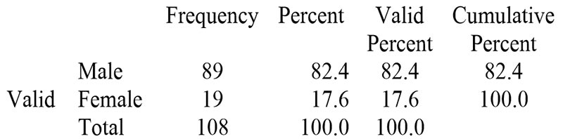
Table 2: Frequency and percentage of age
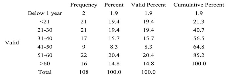
Table 3. Frequency and percentage of clinical information
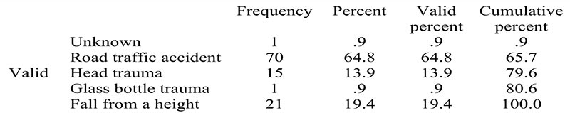
Table 4. Frequency and percentage of common findings

The study evaluated 108 patients who had head computed tomography scan from January to December 2022. The majority of the cases were males (n=89) (82%), while females were (n=19) (17.6%) (Table 1). The majority were within the age of 51-60 years (n=22) (20.4%), followed by those within the age range of 21-30 years (n=21) (19.4%) and <21 years (n=21) (19.4%) and the least were patients below age 1 year (n=2) (1.9%) as seen in Table 2 below. Road traffic accident was the most common clinical indication for CT scan of the head, (n=70) 64.8%, followed by fall from a height (n=21) 19.4%, blunt head trauma (n=15) 13.9% and the least were glass bottle trauma and unknown indication (n=1) 0.9% (Table 3).
With regards to the common brain CT findings, there were hemorrhages (n=64) 52.1%, skull fractures (n = 23) 18.6%, hematomas (n=19) 15.5%, other injuries (n = 8) 6.5%, pneumo-cephalus (n = 7) 5.4% and cerebral edema (n=2) 1.6%. Out of the 108 patients, intra-cerebral bleeding was the highest (n=43) 35%, followed by haemo-sinus (n=21) 17.1% and the least were base of skull fracture, cerebral edema, epidural hematoma and occipital bone fracture with (n=2) 1.6%; subarachnoid haemorrhage and cephal-hematoma were (n=4) 3.3%, subdural hematoma was (n=9) 7.3%, scalp injury was (n=5) 4.1%, temporal bone fracture and orbital injuries were (n=2) 1.6% and frontal bone fracture was (n=16) 13%.
Discussion
The demographic description of cases included in this study showed that majority of the cases were in their second, third, fifth and above sixth decades of their life, with the highest value in the fifth decade of their life, which is 20.4% of the study population. Males were higher in number compared to females. This demographic data is in agreement with the findings of similar studies conducted by Ohaegbulam et al7 (2011) in a medium-sized city (Enugu, Nigeria) and by Ogolodom et al8-9 (2019) in a large city (Port Harcourt, Nigeria). According to Ohaegbulam et al., these categories of people are the most active and productive group in the society and appears to be more prone to both occupational and social risks, while Ogolodom et al. in their study, attributed their findings to the fact that people of this age group are commonly involved in the consumption of hard drugs, cultism, militancy activities and disobeying of traffic rules and regulations, especially the male population. Hemorrhage was the common finding in this study with Road traffic accident as the most common clinical indication. This is however contrary to similar studies by Ugwuanyi et al10 (2020) in Enugu and Haghighi et al11 (2014) in Taba, Shiraz. According to Ugwuanyi et al (2020), out of 300 cases, normal brain CT finding 39% (N = 117) was the most common outcome in their study, despite positive clinical indication to perform the CT scan. According to Haghighi et al (2014) finding, out of 167 patients data included in their study, 147 (88.02%) cases were normal findings, while the abnormal findings accounted for only 20 (11.98%). Similar to our finding, Ohaegbulam et al (2011) in their study, reported 80.1% of abnormal CT findings with the remaining 19.9% been unremarkable. CT is widely available and remains a useful tool for visualizing fracture and bleeding. In this present study, with the clinical indications being mostly road traffic accidents and brain CT findings being more of hemorrhages and fractures, justify the CT scan requests.
Conclusion
Computed Tomography scan is a very useful tool in the evaluation of patients with head injury. The study shows that more males are involved with head injuries than females and that the most common cause of head injury in Umuahia, Abia State is road traffic accident with intra-cerebral haemorrhage being the commonest CT finding.
References
- Kristin M, Johnson D O. The hazard of stopping a brain in motion: Evaluation and classification of traumatic brain injury. AMA J Ethics 10(2008): 516-520
- Hydera A A, Wunderlich C A, Puvanachandraa P, et al. The Impact of traumatic brain injury. A global perspective.Neuro rehabilitation 22(2007) : 341-353.
- Khadka B .Deka P K, Karki A. Role of CT (Computed Tomography) in head injury. JMMIHS 2 (2016): 341-353.
- Mebrahtu-Ghebrehiwet M, Quan L H, Andebirhan T. The profileof CT Scan findings in acute head trauma in Orotta Hospital Asmara Eritrea . J Eritrean MedbAssoc 4 (2009): 5-8.
- Mutch C A.Talbott J F, Gean A. Imaging evaluation of acute traumatic brain injury. Neurosurgery Clin N Am 27(2016) 409-439).
- Hayford K A, Abubakari B A, Emmanuel A, Samson A, Patricia A . Computed Tomography findings of Traumatic Brain Injury in patients with Head Trauma presenting at the Tamale Teaching Hospital, Ghana. Journal of Radiology and Clinical Imaging 2 (2019) : 024-032.
- Ohaegbulam SC, Mezue WC, Ndubuisi CA, et al. Cranial computed tomography scan findings in head trauma patients in Enugu, Nigeria. SurgNeurolInt 2 (2011): 182.
- Ogolodom MP, David LK, Erondu OF, et al. Patterns of traumatic head injury among patients that underwent craniofacial computed tomography scan in Port Harcourt metropolis, Rivers state, Nigeria. Health Sci J 13(2019): 1–8.
- Ogolodom MP, Mbaba AN, Abam R, et al. Magnetic resonance imaging findings in patients presenting with headache in Port Harcourt, Rivers State, Nigeria. J Biomed Sci 8(2019): 3.
- Ugwuanyi DC, Sibeudu TF, Irole CP, Ogolodom MP, Nwagbara CT, Ibekwe AM, Mbaba AN. “Evaluation of Common Findings in Brain Computed Tomography (CT) Scan: A Single Center Study” AIMS Neuroscience Press.7(2020):3,311-318.
- Haghighi M, Baghery MH, Rashidi F, et al. Abnormal findings in brain CT scans among children. J CompPediatric 5 (2014): e13761.